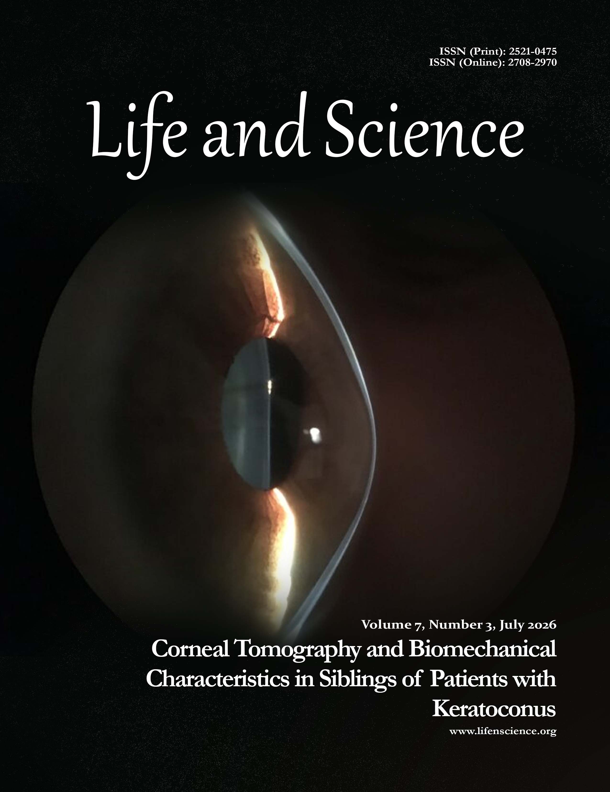
Corneal Tomography and Biomechanical Characteristics in Siblings of Patients with Keratoconus: A Cross-Sectional Study
DOI:
https://doi.org/10.37185/LnS.1.1.1050Keywords:
Corneal Diseases, Corneal Topography, Early Diagnosis, Keratoconus, Risk Factors, TomographyAbstract
Objective: To assess and compare corneal tomographic and biomechanical characteristics between siblings of
patients with keratoconus and healthy siblings of individuals without ocular pathology, aiming to detect early
subclinical changes indicative of increased KC risk.
Study Design: Prospective, observational, cross-sectional study.
Place and Duration of Study: This study was conducted at the Armed Forces Institute of Ophthalmology (AFIO), Pak Emirates Military Hospital (PEMH), Rawalpindi, Pakistan, from 21st August 2024 to 21st February 2025.
Methods: A total of 28 participants (14 in each group) were recruited. Group A included biologically related siblings of Keratoconus patients; Group B consisted of healthy siblings of individuals without ocular disease. Corneal tomography parameters (K1, K2, Kmax, Front Elevation Thinnest Pachymetry, Back Elevation Thinnest Pachymetry, and BAD-D) were measured using Pentacam. Biomechanical parameters, including CBI and TBI values, were assessed via Corvis® ST. Best Corrected Visual Acuity (BCVA) was recorded in logMAR. Data were analyzed using SPSS v25. t-tests and Mann-Whitney U tests were used based on normality, with P < 0.05 considered statistically significant. Effect sizes were calculated.
Results: K2 and Kmax were significantly higher in KC siblings (P < 0.05). D value, Front Elevation Thinnest Pachymetry, and Back Elevation Thinnest Pachymetry also showed significant differences (P < 0.001). CBI and TBI values were significantly higher in the KC sibling group compared to normal siblings (P < 0.001). No significant differences were observed for K1 or logMAR BCVA.
Conclusion: Siblings of KC patients demonstrate early tomographic and biomechanical corneal changes, despite being clinically asymptomatic. These findings support the value of advanced imaging for early detection and risk stratification in high-risk populations.
How to cite this: Machlovi SI, Fawad A, Shah AA, Sohail A, Shabbir A, Mehboob N. Corneal Tomography and Biomechanical Characteristics in Siblings of Patients with Keratoconus: A Cross-Sectional Study. Life and Science. 2026; 7(3): 278-284. doi: http://doi.org/10.37185/LnS.1.1.1050
Downloads
Published
Issue
Section
License
Copyright (c) 2026 Saira Ishaque Machlovi, Aisha Fawad, Aizaz Ali Shah, Azka Sohail, Amina Shabbir, Nimra Mehboob

This work is licensed under a Creative Commons Attribution-NonCommercial 4.0 International License.